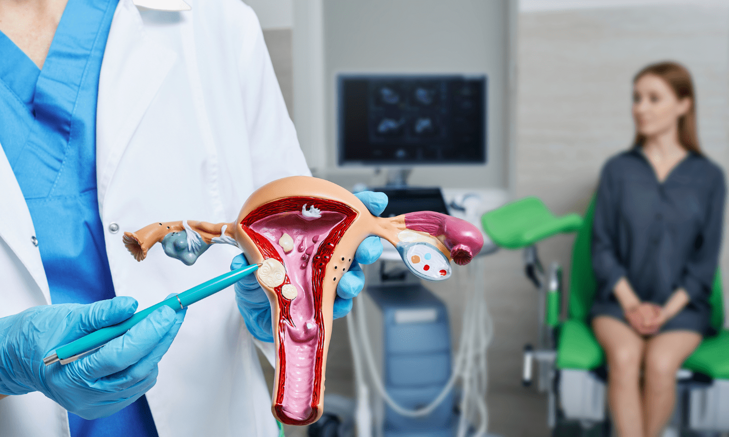
How Are Uterine Fibroids Diagnosed?
Have you ever wondered about those mysterious, uninvited guests called uterine fibroids? They're much more common than you might think. In fact, many women have them without even realizing it! But what are they, and how are they diagnosed? Grab a cup of coffee, get comfortable, and let's dive into this together.
Understanding Uterine Fibroids
Uterine fibroids, or simply fibroids, are non-cancerous growths that develop in or on the uterus, often during childbearing years [1]. Think of them as unwelcome squatters taking up space in your uterus. They vary in size, shape, location, and number. Some are as small as an apple seed, while others can grow as large as a grapefruit!
What are the symptoms of fibroids, you ask? Women may experience heavy menstrual bleeding, prolonged periods, pelvic pressure or pain, frequent urination, or even difficulty getting pregnant. However, some women with fibroids may not have any symptoms at all, which is why regular check-ups are so important.
How Are Uterine Fibroids Diagnosed?
Now let's get to the heart of the matter: how are these fibroids diagnosed? Picture this - you are at your gynecologist's office. You've described your symptoms, and fibroids are suspected. Where do we go from here?
The first step usually involves a routine pelvic exam, similar to what you'd experience during a regular check-up [2]. Your doctor will check the size of your uterus, just like they're sizing up an invisible intruder. If it feels enlarged, fibroids could be the reason.
Imaging Tests for Diagnosing Uterine Fibroids
But a physical exam alone can't give the full picture. That's where imaging tests enter the scene, much like super-detectives with X-ray vision. An ultrasound, often the first imaging test used, works like sonar, using sound waves to detect and create an image of the fibroids.
Sometimes, fibroids play hide and seek, and more specialized tests are needed. One such test is the MRI (Magnetic Resonance Imaging), a powerful tool that can capture detailed images of the size and location of the fibroids [3]. Other tests, like Hysterosonography, Hysterosalpingography, and Hysteroscopy, use a combination of fluids and imaging to visualize the uterus and fallopian tubes more clearly.
Ready to Take the Next Step?
And there we have it! Uterine fibroids might be silent invaders, but with a combination of physical examination and cutting-edge imaging tests, they can't stay hidden for long. The key is early detection and consultation with your doctors. At Indiana Vascular, we extend our expertise to guide you through this crucial diagnostic journey. Your health deserves the spotlight, and early detection is the torchbearer. Schedule an appointment with us today, and let's unveil the mysteries together, laying a solid foundation for your well-informed healthcare journey.
Sources
Cleveland Clinic. Uterine Fibroids: Overview & Facts. Retrieved July 7, 2023, from https://my.clevelandclinic.org/health/diseases/9130-uterine-fibroids
National Institute of Child Health and Human Development. How are uterine fibroids diagnosed? Retrieved July 7, 2023, from https://www.nichd.nih.gov/health/topics/uterine/conditioninfo/how-diagnosed
University of California San Francisco Health. Diagnosis: Fibroids. Retrieved July 7, 2023, from https://www.ucsfhealth.org/conditions/fibroids/diagnosis

